Transmission Electron Microscopy
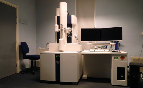
FEI Tecnai T12 (with EDX) and Hitachi HT7800 (with tomography) - click here to find out more.
The BIU is equipped with a wide range of state of the art microscopes and associated equipment to support research and diagnostic imaging with a wide variety of different imaging modalities; light, electrons and X-rays.
FEI Tecnai T12 (with EDX) and Hitachi HT7800 (with tomography) - click here to find out more.
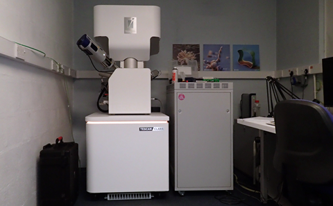
Tescan Clara with serial block face imaging and EDX - second system coming soon - click here to find out more.
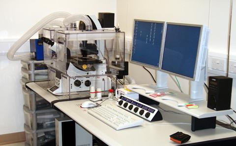
Leica SP5 and Leica SP8 laser scanning confocal microscopes on Leica inverted microscope frames and LaVision Ultramicroscope II light sheet microscope - click here to find out more.
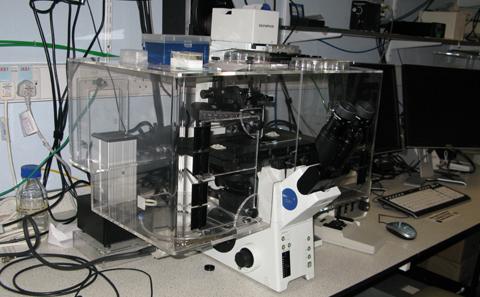
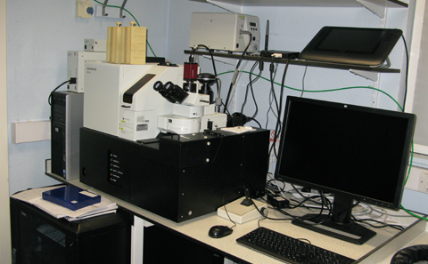
Olympus IX81 fully automated, inverted phase contrast and fluorescence system - click here to find out more.
Olympus dotSlide manual brightfield slide scanners (x2) and Olympus VS110 high throughput slide scanner with fluorescence and phase contrast imaging - click here to find out more.
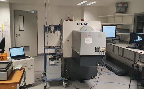
A Milabs whole animal microCT and optical imaging system and Nikon Medex 1 and Medex 2 microCT instruments designed for X-ray histology / soft tissue / low contrast imaging - click here to find out more.
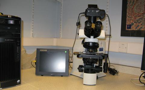
A range of Olympus, Zeiss and Nikon compound and dissecting photomicroscopes with brightfield, darkfield, phase contrast, polarising, fluorescence and DIC imaging modalities - click here to find out more.
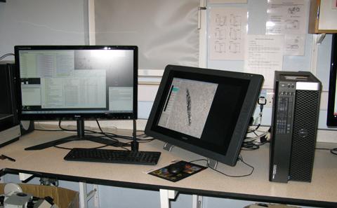
Very high end graphics workstations with touchscreen monitors and a wide range of 2D and 3D image processing and analysis software linked to a multiterabyte network filestore - click here to find out more.
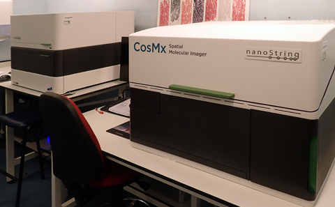
Bruker-Nanostring CosMx and GeoMx instruments with AtoMx online analysis platform, 10x Genomics Visium Cytassist. CosMx and GeoMx instruments are run as a service by BIU - click here to find out more.

A second serial block face SEM system, STED super resolution system, Cryo-fluorescence Tomography system - click here to find out more.