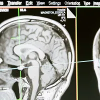
Our advanced three-dimensional imaging and computed tomography (CT) capabilities are based at the µ-VIS X-ray imaging centre. We host a range of different sized µ-VIS (mu-VIS) scanners which work by taking thousands of X-ray views to build up a 3D image of an object.
We also offer 3D X-ray histology (XRH) with our partners at the Biomedical Imaging Unit.
Our services
We provide a holistic approach to advanced 3D-imaging, through our scanning equipment, data analysis capabilities and experienced research staff to support every stage of a project. We welcome collaborations and offer a range of services to external academic and industrial institutions. Typical applications include:
production of images
image analysis (sample visualisation and measurement)
defect detection
actual-nominal comparison
assembly analysis
FE model generation (meshing)
extraction of STL models
We also run introductory training sessions and host workshops on specialist topics.
We have 10 scanners across our 2 sites. They can handle sample sizes with imaged volumes up to 1.5 x 1 x 1m, and spatial resolutions down to approximately 200 nm. We host 6 of the scanners at this location:
Diondo d5 - walk-in scanner
Custom 450/225 kVp Hutch - for specimens up to 100kg
Modified 225 kVp Nikon/Xtek HMX - for specimens less than 300 mm
Zeiss 160 kVp Versa 510 - for specimens in contained in-situ rings
Gatan XuM/Zeiss Evo MA25 - for specimens 1-2mm and below
Nikon/Xtek 160 kVp Benchtop - for training and education
Diondo d5
This is our latest walk-in scanner will band e available to internal and external users from autumn 2022. It is funded by the ESPRC as part of the The National X‑ray Computed Tomography facility.
300 kVp and 450 kVp X-Ray sources
resolutions: 300 kVp source, 450 kVp source
3000 x 3000 FP detector (active area 417 x 417mm)
scanning modes: horizontal and vertical field-of-view expansion and cropping, helical scanning, linear translation laminograpgy
scattering compensation
imaging volume in excess of 1x1x1.5 m
200 kg rating sample stage
temperature controlled
user labyrinth and large 5 x 3.5 x 3.5 m enclosure for large specimens and/or complex in situ testing
Custom 450/225 kVp Hutch
This is best for specimens whose dimensions range from a few millimetres in cross-section up to 1 x 1 x 1.5 m and weigh up to 100 kg.
20 - 225 kVp and 100-450 kVp X-Ray sources
resolutions: ~3µm at low kV, 50µm at 450kV
FP and CLDA detectors
panel shift system
up to 1x1x1.5 m imaging volume
100 kg rating
temperature controlled
large user labyrinth
Modified 225 kVp Nikon/Xtek HMX
This is perfect for small specimens of less than 300 mm in height.
three configurations
transmission (~1µm spot limit, low flux)
standard reflection (~3µm spot, 'normal' flux)
rotating target (~10µm spot limit, x3-5 flux)
2 x 2k flat panel detector
samples to ~300mm and 50kg
robotic sample exchange (~150mm height limit)
Zeiss 160 kVp Versa 510
This allows for imaging of large or small specimens in contained in-situ rings which is useful for biomedical imaging, carbon fibre reinforced polymer (CFRP) and polymer imaging.
30 -160 kVp
2048 x 2048 pixel detector
multiple magnification objectives up to 40x
0.7 µm true spatial resolution
phase-contrast imaging modes applications.
Gatan XuM/Zeiss Evo MA25
This is used in the life sciences for the analysis and reconstruction of biological specimens.
optimised for 4-12kV operation
selection of targets (bulk and thin), ~200nm resolution capabilities
phase enhanced contrast
1340x1300 camera
samples 1-2mm and below
Nikon/Xtek 160 kVp Benchtop
This scanner is available for training and workshops.
30 -160 kVp
reflection source, ~3µm spot size limit
1248x1248 flat panel detector
samples to ~90mm diameter, 5kg
Scanners at our partner site
Our 4 specialist histology scanners are hosted at the hospital:
Nikon XTH225ST XRH 3D X-ray histology scanner
Nikon Med-X (alpha)
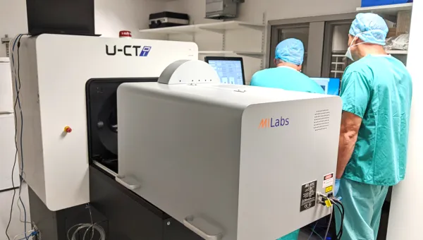
MILabs U-CT 3D/4D optical imaging / microCT system
SkyScan 1176 in-vivo CT scanner
Research
Our 3D X-ray scanners provide a non-destructive way of looking inside materials and objects in great detail. This is a huge advantage for a wide range of research areas that need to study objects which are too unique, delicate or complicated to take apart. We’ve worked closely on a diverse range of applications with the aeropsace and automotive industries, museums, accident investigators and the rail industry.
Research applications
Our scanners have been used to:
inspect the internal geometry of electrothermal thrusters for use in small spacecraft
study fatigue in rail axle bearings
measure the active surface area of different grades of reticulated vitreous carbon for use in electrochemical sensors and batteries
investigate cracks in nickel based single crystal superalloys used in advanced turbine engines
study Roman coins without breaking the pots they were buried in
virtually excavate the wreck of the Rooswijk, which sank off Kent in 1740
Visit our YouTube channel to watch 3D rendering movies of some of our work.
Our team includes directors Professor Ian Sinclair and Dr Mark Mavrogordato, together with experienced beam scientists and over 40 academic staff covering a wide range of expertise.
μ-VIS X-Ray Imaging Centre,
Eustice building (B5),
Faculty of Engineering and Physical Sciences,
University of Southampton,
Highfield Campus,
Southampton, SO17 1BJ