Our diagnostic services include:
- transmission electron microscopy of a variety of biopsy types for pathological conditions that are still only diagnosable by ultrastructural analysis, especially renal disease
- scanning electron microscopy of hair samples
- x-ray microanalysis of pathological inclusions using scanning or transmission electron microscopy
Primary Ciliary Dyskinesia (PCD) diagnostic service
We are one of 3 national referral units funded by the NHS National Commissioning Group for the specialist diagnosis of PCD through combined light and electron microscopy approaches. PCD is a syndrome of rare hereditary diseases that affect the structure, function and behaviour of cilia in the airways. It impairs lung clearance and produces lung disease. Other biological processes that involve cilia/flagellae, from patterning of the early embryo through to fertility in adults can also be affected.
Clinical diagnosis of PCD includes:
- clinical history and referral for persistent chronic respiratory infection
- collection of ciliated epithelia cells by brushing the nasal passages
- high speed video microscopy of ciliated nasal epithelial cells to reveal defects in ciliary beat frequency, stroke pattern and coordination
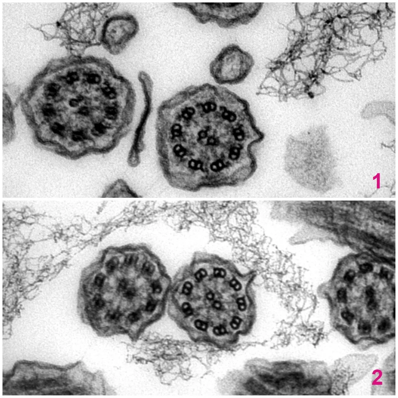
- transmission electron microscopy of ciliated nasal epithelial cells to examine the ultrastructure of individual cilia and locate missing or misarranged ciliary components
Visit the Southampton Children's Hospital PCD diagnostic service
Accreditation
Within University Hospital Southampton, we are part of the wider Cellular Pathology division which holds ISO 15189 accreditation.

- A transmission electron microscope image of a single healthy cilium in cross section. The axonemal structure of microtubules is correct and the dynein arms are present.
- A transmission electron microscope image of a single cilium in cross section from a patient with Primary Ciliary Dyskinesia. The axonemal structure of microtubules is correct however the dynein arms are absent.